Are mammograms safe? It’s a question that many have asked. New research questions the effectiveness of mammograms. Because of this, the American Cancer Society changed its recommendation on mammography in October 2015. The ACS had recommended annual mammograms beginning at age 40. Now that we know more about the limitations and risks involved, the ACS recommends that mammograms shouldn’t begin until age 45 and should only be every other year at age 55.
There are alternatives to mammography. Thermography offers a non-invasive method to examine the physiology of the body. This is different from mammograms and ultrasound, which examine the structure of the body.
Thermography detects subtle variations in skin temperature using an infrared camera in a temperature-controlled room. This process offers insights into what may be happening below the surface of the skin. Humans give off energy in the form of heat. An infrared camera is heat-sensitive. A mammogram emits ionizing radiation through your compressed breast tissues. Thermography differs in that you radiate energy towards the camera. Nothing is passed through your body.
Cancer feeds on your own blood supply. Thermography detects the higher heat the results from the development of vascularity (angiogenesis) that feeds cancer. When cancer grows, the normal cell-death mechanism, known as apoptosis, stops. The cell doesn’t die and continues to grow. Since the early process is at a cellular level, a solid mass hasn’t formed yet. It typically takes a few years for a cancerous tumor to grow large enough to be detected on a mammogram.
The chart below was developed from Dr. Michael Retsky’s cancer-growth research showing that the possible observation times for a mammogram to find a tumor are near the end of the tumor’s growth, which does not constitute early detection. His research found that breast cancer typically doubles in volume in about 100 days. Since mammography is usually able to find breast tumors at approximately 1 cm, he estimates the usual time to detect breast cancer is at 30 doublings (of 100 days each)-a total of 8 years. He concludes that “the possible observation times in breast cancer is limited to between the 30th and 40th doublings or at most the last 25% of the growth history of a tumor.”
| 90 days | 2 cells |
| 1 year | 16 cells |
| 2 years | 256 cells |
| 3 years | 4,896 cells |
| 5 years | 1,048,576 cells |
| 6 years | 16,772,216 cells |
| 7 years | 268,435,456 cells |
| 8 years | 4,294,967,296 cells |
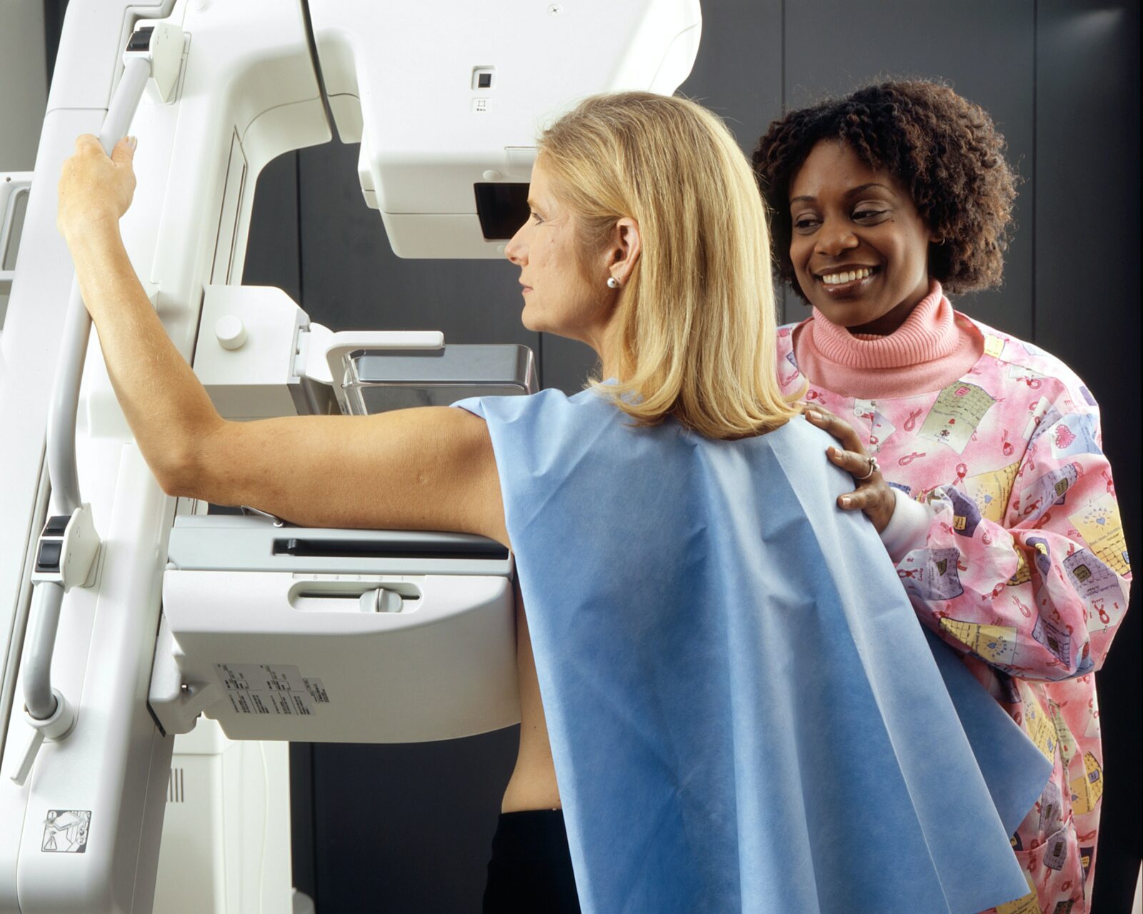
Mammography
Mammograms first caused concerns due to a large number of false positives in addition to overdiagnosis and overtreatment. Some studies found that mammograms did not improve overall breast cancer mortality rates most likely because they don’t offer true early diagnosis. Most often breast cancers are found in the upper outer area of the breasts, in between the breast tissue and the armpit, which cannot be visualized on a mammogram.
Mammograms have an average sensitivity of 80% in women over 50, which drops to 60% in women under 50. Using hormones decreases the sensitivity of mammograms. In addition, mammograms do not differentiate between a solid tumor and fluid-filled cyst or calcification. Thus, women who have scar tissue or dense or fibrocystic breasts get called back to have a repeat mammogram because of difficulties reading the scans. This increases radiation exposure. Still, the National Cancer Institute reassures women that the benefits of mammography outweigh the risks. But repeated X-ray exposure can cause cancer.
Thermography
Thermography is a reasonable alternative for women who want to avoid the radiation exposure from a mammogram, for those who have implants, or for women who have had other breast surgeries resulting in scar tissue. It is also a great option for women who are considered high risk, are taking hormones, are younger, or have dense breasts. Thermography has no harmful side effects, so it can be used frequently without concern.
According to the American College of Clinical Thermography, thermography can detect abnormalities of the female breast and can also examine breast tissue in men. Another advantage is that the entire chest is examined, and there is no compression of tissue, which can sometimes spread cancer cells. Thermography can also monitor treatment effectiveness and can distinguish between benign and malignant tissue in women with fibrocystic breasts.
Over 800 peer-reviewed articles support the effectiveness of thermography, and the process was approved by the FDA as an adjunctive test to mammography in 1982.
A 2003 study indicated “Thermography offers a safe, noninvasive procedure that would be valuable as an adjunct to mammography in determining whether a lesion is benign or malignant with a 99% predictive value.”
A study published in 2008 by The American Society of Breast Surgeons concluded that DITI (digital infrared thermal imaging, or thermography) was a valuable adjunct to mammography and ultrasound especially in women with dense breast parenchyma [tissue] because of its 97% sensitivity.
In 2013, researchers Kolaric et. al. found thermography to have the probability of a correct finding in 92% of cases. They concluded that “breast cancer remains the most prevalent cancer in women and thermography exhibited superior sensitivity. We believe that thermography should immediately find its place in the screening programs for early detection of breast carcinoma, in order to reduce the suffering from this devastating disease.”
According to women’s health specialist, Dr. Christine Horner, thermography can “detect breast cancers much earlier than any other available technology. Because blood vessels ordinarily start to grow before any other significant changes and tumor growth, a thermogram can ‘see’ these abnormal physiological processes as early as 5-10 years before cancer can be seen by a mammogram, MRI, or ultrasound or felt by a physical exam. What is most exciting is that when these abnormal processes are caught this early, they are reversible.”
The ACCT says thermal imaging should be “viewed as a complementary, not competitive, tool to mammography and ultrasound” that can increase the effectiveness of those two structural tests by identifying patients having the highest risk level.
The International Academy of Clinical Thermography says that thermography is not a replacement for mammography because “there is no one test that can detect 99-100% of all cancers.” In addition, thermography and mammography “are ‘looking’ for completely different pathological processes” because one tests physiology and the other tests anatomy. Still, thermography is more sensitive than mammography. However, mammography can detect some slow-growing, non-aggressive cancers better than thermography.
Breast cancer detection is a multifaceted issue that requires an individualized approach. You should do your own research and discuss your options with your doctor. Both thermography and mammography are good detection tools. Which one you use may depend on factors like age, disease status, history of disease, density of breast tissue, type of cancer, and more. Remember that thermography, mammograms, or breast exams cannot diagnose cancer. If something suspicious is found on a mammogram, or by ultrasound, breast exam, or thermography, the definitive diagnosis can only be done by biopsy.